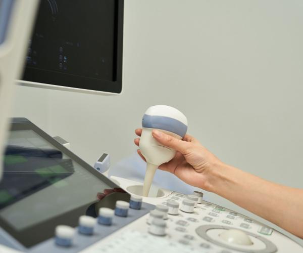
Beast Ultrasound
Breast ultrasound is a non-invasive diagnostic test that uses high-frequency sound waves to create images of the breast tissue and surrounding structures. It is a safe and painless method that does not involve radiation, making it suitable for women of all ages, including pregnant women.

When Is a Breast Ultrasound Performed?
What Can a Breast Ultrasound Detect?
Cysts
Round or oval fluid-filled areas.
Fibroadenomas
Benign tumors, common in younger women.
Lipomas
Benign fatty tissue tumors.
Inflammation
Μαστίτιδα, απόστημα.
Cancer
Solid masses with irregular borders or vascularization.
Lymph Node Changes
Enlargement or structural alterations.
When Is a Breast Ultrasound Recommended?
If you notice a palpable mass or abnormality in the breast.
If you experience symptoms such as
- Breast pain
- Nipple discharge
- Change in breast size or shape
- Redness or thickening of the skin
If there is a family history of breast cancer.
If a mammogram reveals a vague or suspicious finding.
Psarras - Reserch Medical S.A.
Advantages of Breast Ultrasound
- Non-invasive
No surgical intervention required. - Radiation-free
Completely safe. - Accurate in dense breast tissue
Ideal for younger women. - Guided biopsy capability
Useful for tissue sampling. - Quick and painless
Takes approximately 15–30 minutes.
Limitations of Breast Ultrasound
Does not replace mammography because:
- Cannot detect microcalcifications (early sign of cancer).
- Does not provide a complete map of breast tissue like a mammogram.
Στο ΨΑΡΡΑΣ-Ερευνητικό Ιατρική ΑΕ διενεργούνται οι παρακάτω
εξετάσεις Μαγνητικής Τομογραφίας:
Άνω κοιλίας
Κάτω κοιλίας
Οπισθοπεριτοναϊκού χώρου
Εγκεφάλου
Σπλαχνικού κρανίου
Λιθοειδών οστών
Οφθαλμικών κογχών
Υπόφυσης
Παραρρινίων κόλπων
Μαλακών μορίων
Τραχηλικής χώρας
Θώρακος
Αυχενικής Μοίρας Σπονδυλικής Στήλης
Θωρακικής Μοίρας Σπονδυλικής Στήλης
Οσφυϊκής Μοίρας Σπονδυλικής Στήλης
Ιεροκοκκυγικής Μοίρας Σπονδυλικής Στήλης
Αρθρώσεων (ώμου, αγκώνος, πηχεοκαρπική άρθρωση και άκρα χείρα, ισχίου, γόνατος, ποδοκνημική άρθρωση)
Contact Us
Have questions? Get in touch!
Error: Contact form not found.
Contact
Send us your message and we will contact you as soon as possible.


