THE ROLE OF HIGH-RESOLUTION ULTRASOUND IN THE DIAGNOSTIC APPROACH TO DUCTAL CARCINOMA IN SITU (DCIS) OF THE BREAST
Eir. Georgiou¹, K. Psarras¹, N. Kritikos¹, D. Daskalopoulou², R. Angelatou¹
1 Breast Imaging Department, GAONA “Agios Savvas”, Athens
2 Cytology Department, GAONA “Agios Savvas”, Athens
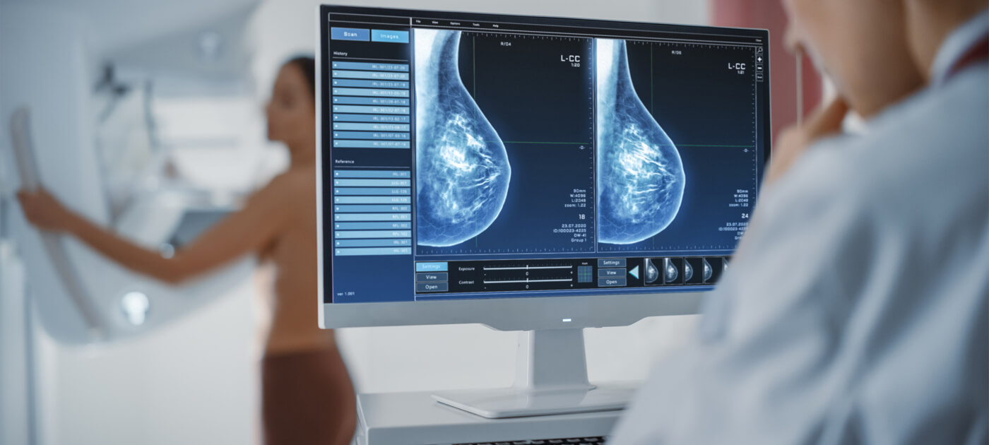
Ductal carcinoma in situ (DCIS) of the breast is often visualized mammographically as a non-palpable mass with calcifications. The aim of this study, conducted at GAONA “Agios Savvas,” was to evaluate the capability of high-resolution ultrasound in detecting DCIS by describing the imaging characteristics of the lesions, and to highlight the significance of ultrasound-guided fine-needle aspiration (FNA) in histological identification.
The study included 75 cases of DCIS managed in the breast imaging department. Among these, 23 cases (Group A) were diagnosed solely using ultrasound, while the remaining 52 cases (Group B) were diagnosed mammographically or during clinical examination. All cases were histologically confirmed via ultrasound-guided FNA, and the results were compared with those of open biopsy.
Ultrasound findings in Group A revealed cystic or solid lesions in 18 patients (80%). The mean age of Group A was significantly higher than that of Group B (60 years vs 51 years). Tumor size in Group A was about half that of Group B, and intraductal extension was also smaller. Most tumors were of low-grade malignancy. Additionally, tumors smaller than 5mm were predominantly low-grade.
The conclusion is that the growing use of high-resolution ultrasound along with ultrasound-guided interventional procedures has significantly increased the detection rate of breast cancers that are not visible on mammography. Most of these are DCIS or small invasive carcinomas, and when detected via ultrasound, they are typically low-grade in malignancy.