Select your language

Ultrasonic - Triplex

Ultrasonic - Triplex

Ultrasonography is a medical imaging method that produces images of parts of the human body using ultrasound.

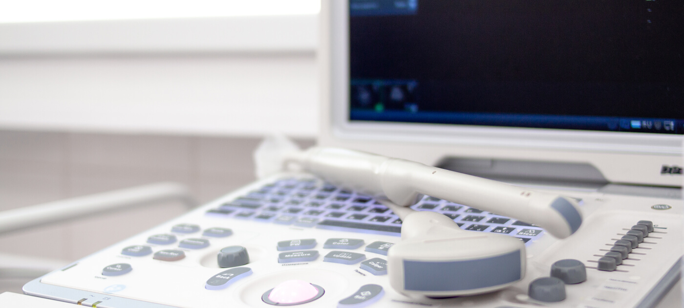
Our centre has the state-of-the-art LOGIQ S8 XDclear ultrasound scanner from General Electric.

It is a machine with a computer-like architecture that receives signals from a transceiver, processes them and converts them into black-and-white or colour images.

The ability of ultrasound to easily, timely and reliably discover lesions along with the ability to document their benignity or malignancy, makes it extremely important in the field of preventive medicine.

    The ultrasound examination performed on the vessels is called TRIPLEX, because the structures of the vessels are checked simultaneously with the blood flow. It is a simultaneous morphological and hemodynamic study. With this test, the doctor sees how the blood flows in the vessel and measures the flow rate.

    In this way it can assess:

  • The flow of the blood in the vessel and its size
  • If there is a stenosis or obstruction of the vascular lumen
  • The changes that the blood flow undergoes due to the constrictions

    A. Peripheral vessels

  • Carotid arteries and vertebral arteries
  • Upper limb arteries
  • Lower limb arteries
  • Upper limb veins
  • Lower limb veins

    Β. Visceral vessels

  • Abdominal aorta
  • Renal arteries
  • Splenopelvic axis