

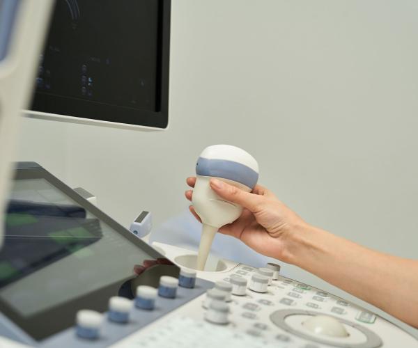
Breast ultrasound is a non-invasive diagnostic test that uses high-frequency sound waves to create images of the breast tissue and surrounding structures. It is a safe and painless method that does not involve radiation, making it suitable for women of all ages, including pregnant women.
When Is a Breast Ultrasound Performed?
Evaluation of a Palpable Mass
– Determines if the mass is solid (potential tumor) or cystic (fluid-filled).
Follow-Up After a Mammogram
– If a mammogram detects a suspicious area, an ultrasound can provide more detailed imaging.
Screening for Younger Women (<40 years)
– Breast tissue in younger women is denser, making mammograms less accurate.
Guidance for Biopsy
– Used to guide the needle for tissue sampling in case of suspicious findings.
Monitoring Benign Tumors
– Used to monitor fibroadenomas or cysts for any changes over time.
Axillary Lymph Node Evaluation
– Detects enlarged or suspicious lymph nodes in the armpit area.
What Can a Breast Ultrasound Detect?
Cysts
Fibroadenomas
Lipomas
Inflammation
Cancer
Lymph Node Changes
When Is a Breast Ultrasound Recommended?
If you notice a palpable mass or abnormality in the breast.
If you experience symptoms such as
- Breast pain
- Nipple discharge
- Change in breast size or shape
- Redness or thickening of the skin
If there is a family history of breast cancer.
If a mammogram reveals a vague or suspicious finding.
Psarras – Research Medical S.A.
Advantages of Breast Ultrasound
- Non-invasive
No surgical intervention required. - Radiation-free
Completely safe. - Accurate in dense breast tissue
Ideal for younger women. - Guided biopsy capability
Useful for tissue sampling. - Quick and painless
Takes approximately 15–30 minutes.
Limitations of Breast Ultrasound
Does not replace mammography because:
- Cannot detect microcalcifications (early sign of cancer).
- Does not provide a complete map of breast tissue like a mammogram.