

Ultrasonic
Ultrasound is a diagnostic imaging technique that uses high-frequency sound waves to create images of the internal organs and tissues of the body.
Our center is equipped with the state-of-the-art LOGIQ S8 XDclear ultrasound machine from General Electric — a highly advanced system known for its precision and clarity.
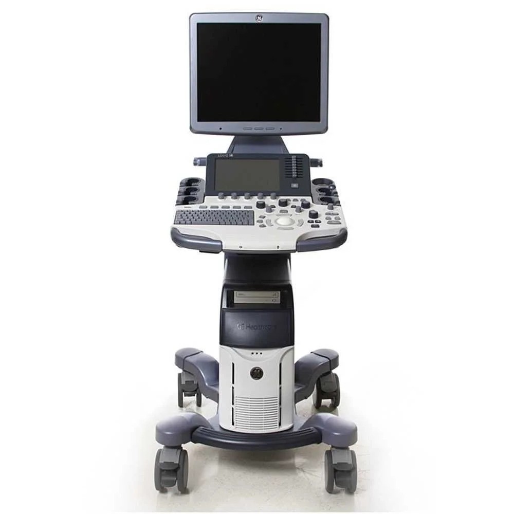
Psarras – Research Medical S.A.
Advantages of Ultrasound
- Non-invasive
No surgical procedure required. - Painless
No use of radiation. - Safe
Suitable for pregnant women and children. - Fast
Provides real-time images.
What an Ultrasound Can Reveal?
- Organ morphology
Thickness, size, and shape of organs. - Pathological conditions
Cysts, tumors, inflammation, and fatty infiltrations. - Blood flow
Evaluation of blood circulation in arteries and veins (using Triplex Doppler). - Structural changes
Hardening, swelling, or tissue damage.
Triplex
Vascular Triplex is a specialized ultrasound examination used to evaluate blood flow in the arteries and veins. It combines three imaging techniques to provide a comprehensive view of vascular health:
Imaging techniques
01
2D (B-mode) Imaging
– Displays the structure and anatomy of blood vessels.
02
Doppler Imaging
– Measures the speed and direction of blood flow within the vessels.
03
Color Doppler Imaging
– Colors the blood flow, showing its direction and intensity.
When Is a Vascular Triplex Performed?
Diagnosis of Vascular Conditions
- Atherosclerosis → Narrowing or blockage of arteries.
- Venous insufficiency → Poor blood return from the veins.
- Thrombosis → Presence of blood clots.
- Aneurysms → Enlargement of blood vessels.
Assessment of Blood Circulation
- Whether blood flow is normal or reduced.
- Whether there is backward flow (reflux).